 图片附件: 98112623.snap.jpg (2011-12-18 15:18, 34.91 KB) / 该附件被下载次数 20
 图片附件: 18174162.snap.jpg (2011-12-18 15:19, 38.79 KB) / 该附件被下载次数 10
 图片附件: 14042777.snap.jpg (2011-12-18 15:20, 29.15 KB) / 该附件被下载次数 18
 图片附件: 55712497.snap.jpg (2011-12-18 15:20, 39.77 KB) / 该附件被下载次数 76
 图片附件: 86822896.snap.jpg (2011-12-18 15:21, 31.27 KB) / 该附件被下载次数 17
 图片附件: 51652739.snap.jpg (2011-12-18 15:21, 25.47 KB) / 该附件被下载次数 12
 图片附件: 57171612.snap.jpg (2011-12-18 15:21, 53.57 KB) / 该附件被下载次数 13
 图片附件: 88233009.snap.jpg (2011-12-18 15:22, 57.1 KB) / 该附件被下载次数 14
 图片附件: 43526812.snap.jpg (2011-12-18 15:28, 43.39 KB) / 该附件被下载次数 2
 图片附件: 46796089.snap.jpg (2011-12-18 15:40, 44.31 KB) / 该附件被下载次数 13
 图片附件: 40325993.snap.jpg (2011-12-18 16:49, 47.25 KB) / 该附件被下载次数 3
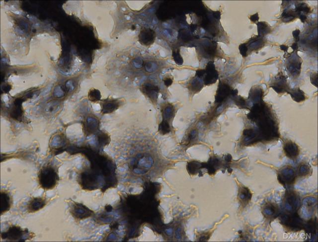 图片附件: 68805870.snap.jpg (2011-12-18 16:50, 43.23 KB) / 该附件被下载次数 3
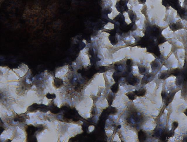 图片附件: 55132809.snap.jpg (2011-12-18 16:50, 43.21 KB) / 该附件被下载次数 4
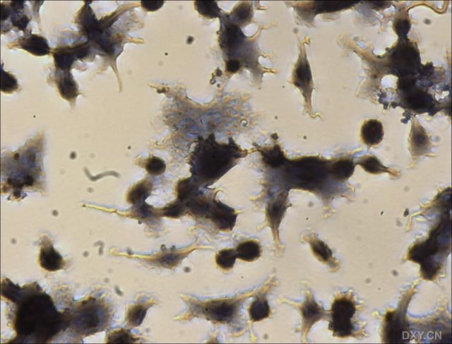 图片附件: 50131196.snap.jpg (2011-12-18 16:51, 41.44 KB) / 该附件被下载次数 3
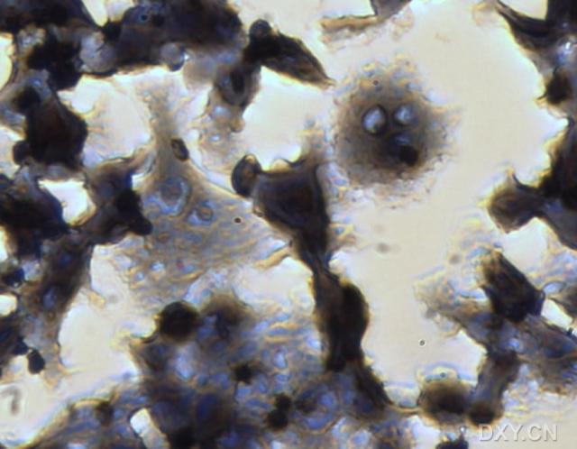 图片附件: 42884347.snap.jpg (2011-12-18 17:11, 37.49 KB) / 该附件被下载次数 7
 图片附件: 86575780.snap.jpg (2011-12-18 17:11, 66.29 KB) / 该附件被下载次数 2
 图片附件: 52753471.snap.jpg (2011-12-18 17:12, 49.12 KB) / 该附件被下载次数 7
 图片附件: 96695642.snap.jpg (2011-12-18 17:12, 45.46 KB) / 该附件被下载次数 3
 图片附件: 88116878.snap.jpg (2011-12-18 17:16, 45.49 KB) / 该附件被下载次数 3
 图片附件: 72425302.jpg (2011-12-18 17:16, 81.12 KB) / 该附件被下载次数 3
 图片附件: 97917722.snap.jpg (2011-12-18 17:17, 53.22 KB) / 该附件被下载次数 4
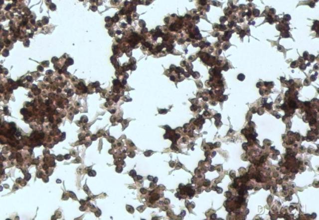 图片附件: 93421383.snap.jpg (2011-12-18 17:17, 56.94 KB) / 该附件被下载次数 0
 图片附件: 76041203.jpg (2011-12-18 17:18, 74.03 KB) / 该附件被下载次数 130
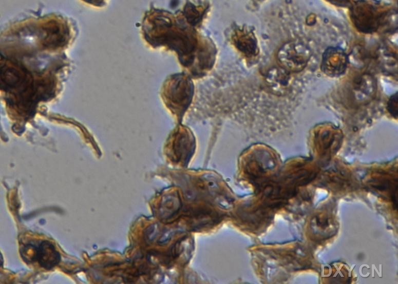 图片附件: 88000588.snap.jpg (2011-12-18 17:42, 62.52 KB) / 该附件被下载次数 8
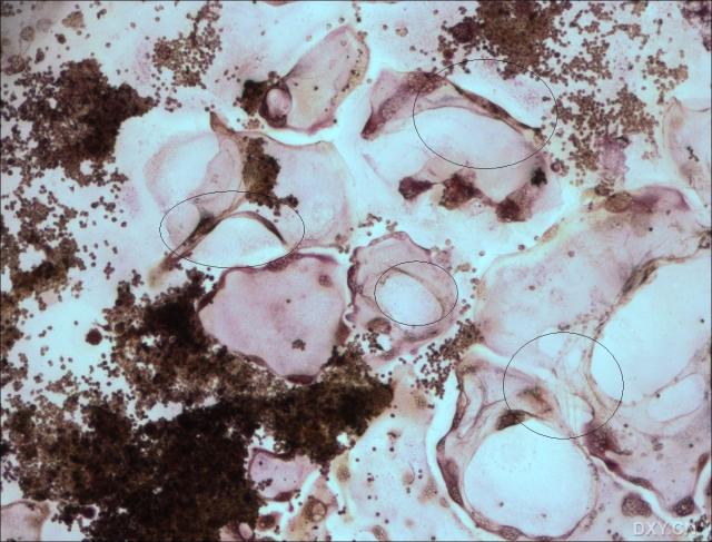
| 欢迎光临 分析测试百科 (http://bbs.antpedia.com/) | Powered by Discuz! 5.5.0 |