我用40000vhr试过了,结果还是不行。我现在怀疑是我的sample提取的问题。
而且我的胶好像并不存在过聚焦的问题,而可能是样品沉淀的问题。
下面我列出了我的2-D protocol,各位战友帮我看看有没有问题?如何改善?谢谢!
I. reagents prepared in advance
1. Tris-buffered sucrose (stored at 4ºC)
Tris 10mM 1.21g
Sucrose 250mM 85.6g
ddH2O to 1000ml, then to pH7
2. lysis solution (stored at -20ºC)
Urea (FW60.06) 8M 19.2g
CHAPS 4% 1.6g
ddH2O to 40 ml
transfer into eppendorff tubes, 1ml / tube, stored at -20 ºC.
3. rehydration buffer (without DTT, IPG buffer)
Urea 8M 12g
CHAPS 2% 0.5g
Bromophenol blue trace
ddH2O to 25ml
loaded into eppendorff tubes, 1ml / tube, stored at -20 ºC
4. DTT (FW=154.2)
Stock 700mg/ml, 4ul /tube, 2.8mg, stored at -20 ºC.
2.8mg / 1ml =18mM
II. sample preparation
1. 10-cm dish with 90% confluence cells.
4 x 10⁵/ml x 10ml cells, cultured in 10-cm dish, 48hrs later cells are collected, with concentration of about 1.2 x 10⁶/ml x 10ml.
2. Operation on ice. Wash with “Tris-buffered sucrose” 3ml, 3 times, suck waste by vacuum.
3. Treat with 250μl lysis solution for 15 min on ice. Add protease inhibitors to lysis solution (5 μl PI / 1 ml lysis solution) prior to use.
4. Scrape the cells with a cell scraper, and transfer cells to eppendorff tube on ice with cut-tip.
5. Sonication on ice: pulse, 10 sec start, 10 sec stop, 4 times. (Amp 16%)
6. Centrifugation: 13,000 rpm, 18°C, for 30 min.
7. Supernatant was collected, protein concentration of which was detected with Bradford assay, and stored at -70°C.
III. strip rehydration in advance
1. Select 13-cm IPG strip, with pH3-10.
2. 250 μl rehydration solution / 13 cm IPG strip.
3. add DTT and IPG buffer into rehydration solution prior to use. One ml rehydration solution with 4μl DTT (2.8mg) + 0.5% pH3-10 IPG buffer .
4. add rehydration solution into Reswelling Tray. Rehydrate strip gels, with the gel side down.
5. overlay strip with cover fluid, about 3 ml.
6. Allow the IPG strips to rehydrate overnight (10–20 h).
IV. IEF (first dimension)
1. Select holders. Put strip (after rehydration) into holder, with gel side up.
2. Use the paper wicks on “+” and “-” sides. Paper wicks must be damp, not wet. Remove excess water by blotting with tissue paper. Put electrodes onto paper wicks.
3. Put the cup on anode side, and use cover fluid to test leakage.
4. Add sample into cup. Sample volume + rehyration buffer + DTT (0.4ul, 0.28mg) + 0.5% IPG buffer =100 ul total volume
5.Overlay the gel with Cover Fluid, 3-4ml.
6. Run programme
IEF running conditions: 20°C, 50 uA/strip
S1 100v 30min step-n-hold
S2 250v 30min step-n-hold
S3 500v 30min step-n-hold
S4 1000v 30min step-n-hold
S5 2000v 30min step-n-hold
S6 4000v 30min step-n-hold
S7 8000v 48,000vhr gradient
S8 30v 10hr step-n-hold
S9 0v 0hr
Ao7 (2014-4-26 15:48:15)
7. take out strip and store it at -70°C.
V. prepare second-dimension gel in advance
1. set up gel casting cassette and use ddH2O to test leakage.
2. prepare light gel stock (0%) and heavy gel stock (20%).
3. cast gel with gradient concentration of 8-16%, using gel mixture instrument.
4. block out the up-side of gel with ddH2O.
5. allow gel to freeze for more than 3 hr.
VI. SDS-PAGE (second-dimension electrophoresis)
1. Preparation in advance.
SDS-equilibration buffer (stored at -20ºC, 10ml/tube)
1.5M Tris –HCl (pH8.8) 6.7ml (50mM)
Urea 72.07g (6M)
Glycerol 69ml (30% v/v)
SDS 4.0g (2%)
Bromophenol blue trace
ddH2O to 200 ml
Note: Do not add SDS from the beginning, because urea and SDS tegother would need a long time to dissolve.
2. Add 100mg DTT into 10ml buffer. Place strip in 5ml buffer, shake 15min, RT, with gel-side up.
3. 250mg IAA / 10ml buffer, 5ml / strip, shaking 15min, RT, gel-side up.
4. prepare “Agarose sealing solution”
Running buffer 100ml
Agarose 0.5g (0.5%)
Bromophenol blue 0.01 g (0.01%)
5. prepare paper marker (5-10ul, Marker 12 in 4x4mm paper)
6. Open cooling system in advance (14ºC).
7. Electrophoresis condition:
1.5mm-thick gels, 15mA/gel for 1 hr, then 35 mA/gel for about 2.5 hr.


最新回复
Ao7 (2014-4-26 15:27:54)
(2)
55107664.snap.jpg
Ao7 (2014-4-26 15:28:22)
(3)
63721939.snap.jpg
Ao7 (2014-4-26 15:28:58)
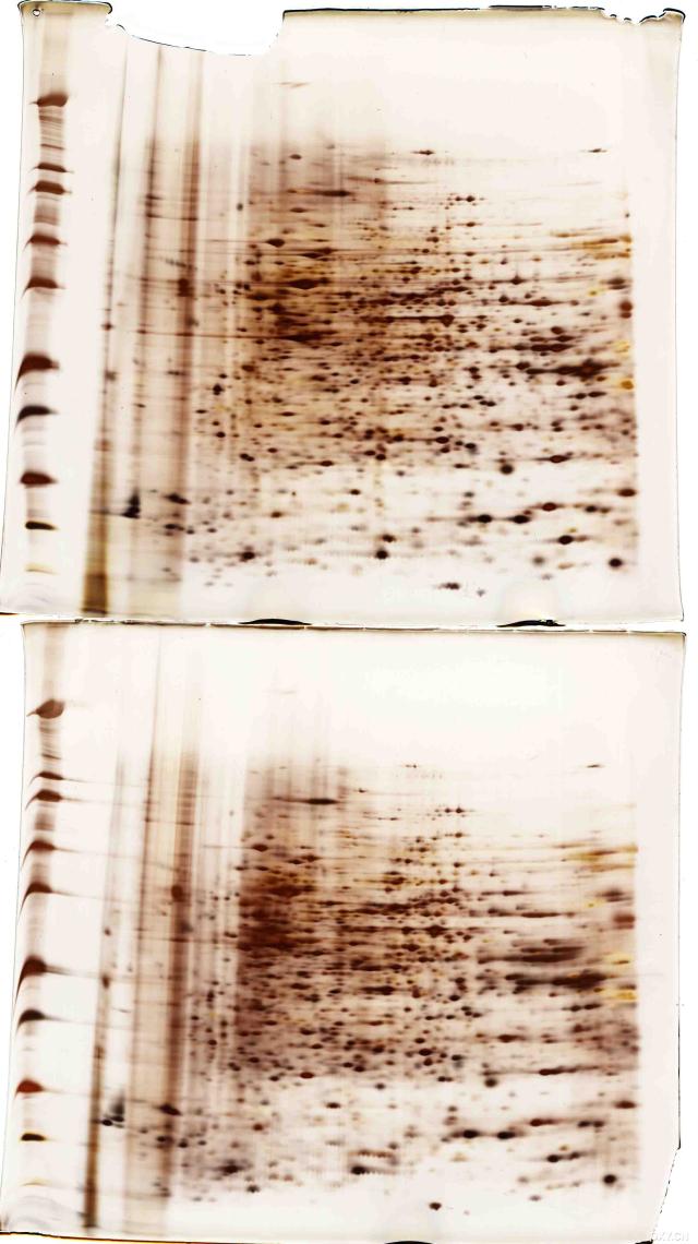
我做的是HepG2细胞的对照组和处理组。由于以前用IPG buffer=0.5%经常跑出像图(2)这样的结果来,于是图(1)我改用IPG buffer=1%,结果感觉还可以。但是用不同的样品用IPG buffer=1%重复了4次,却又出现了类似于图(2)的结果。于是,我又用图(1)相同的sample,但用IPG buffer=0.5%跑了2次,得到了图(3)的结果。很显然,重现性不是太好。
我的一向电泳的电压最高在4000-6000,从未到达80000.
另外,我的是13cm strip,PH3-10,聚焦为5万Vh.
我要向高手请教的是:IPG buffer浓度多少合适?是不是2-D重现性本身就不好?图(2)和图(3)可能是什么问题?如何能改善我的一向电泳的电压又使我的sample不会发生沉淀?
谢谢!我已郁闷得不行了!希望好心人能帮帮忙!再次谢谢!
最好能让我改动一个指标就有明显的改善:)
Ao7 (2014-4-26 15:29:16)
另外,想请教:图(2)和图(3)中在cup边上以及中间两处都有明显的gap,到底是怎么回事?
GE手册说是因为sample不溶造成的,那为什么(1)却没有?图(1)和(3)是相同的sample,放在-80度冰箱中保存的,都只解冻一次。
8s5g (2014-4-26 15:29:58)
虽然2DE的重现性是个问题,但你的结果重现性也未免太差了点。
不知在这几次试验中,你除了IPG buffer外,还改变了什么条件没?
此外,13cm的胶条,你可试试略减小点聚焦伏小时数,试试40000VHr。
Ao7 (2014-4-26 15:30:33)
而图(1)和图(2)是不同的sample, IPG buffer都为1%.用的是cup loading.只是图(1)150ug (18.5ul)+rehydration buffer(包括DTT,IPG buffer)=100ul 总的cup loading 体积。而图(2)则用了150ug(31.5ul),蛋白浓度较稀而已。
为什么会这样?!
请您多多提建议,我是新手,向您学习!
Ao7 (2014-4-26 15:30:57)
我下次会试试小一点的Vhrs,如您所说的4万vhrs.
另外,想请教这是否与盐份或其它致污物有关?图(1)只是运气好而已?所以,为了结果稳定,还要除盐,是吗?
请问有哪些简便而有效的除盐方法?有protocol吗?谢谢!
8s5g (2014-4-26 15:32:20)
=======================================================================
感觉上无论IpG buffer是1%还是0.5%,聚出来的点都还挺好,但点的数量明显不同。
图(3)的左半部分,感觉转移没有转好,丢失了很多点。
此外,是否在样品保存中有问题,导致部分蛋白降解?或者解冻时没有充分溶解就上样了?
2DE的结果差异有时候很难说,这和水化过程、平衡、胶条转移、及SDS胶的配制都相关。
8s5g (2014-4-26 15:41:09)
==============================================================================================================
图(2)明显聚焦不好,此外,蛋白浓度较低会出现这样的现象,我在做时觉得蛋白浓度低3-5ug/ul就是没有蛋白浓度高,如7-10ug/ul的样品跑得好。
但具体原因也不是太清楚。希望其他有经验的战友帮忙解答。
Ao7 (2014-4-26 15:45:10)
谢谢8s4g,我这次重新提取了蛋白,浓度约为10ug/ul,IPG=0.5%,然后打算聚焦4万vhrs.希望可以得到较好的图。
我只是想不通:图(2)和图(3)中在杯(cup)边上以及中间分别有明显的空白带(gap)。GE公司的手册上说致污物可以造成空白。我做了许多次(N>10)2-D,除了少数没有空白带以外,其它的胶在相同的pH位置都有空白带。我做过pH4-7的胶,也在相同pH位置出现。我真的很想知道到底是哪一种因素造成的?可能是哪些致污物造成的?我只有知道了原因才能有的放矢。
其他战友有类似的经验吗?最近通过哪种方式改善的?
我已经困惑了很长一段时间了,对自己都开始没有信心了。还希望各位战友不吝赐教。
先在此谢过!
zzzz (2014-4-26 15:45:34)
我在做时觉得蛋白浓度低3-5ug/ul就是没有蛋白浓度高,如7-10ug/ul的样品跑得好。
--------
这一点我赞同,我的样品上次提的只有3.5ug/ul左右的,效果就很差,几乎点都看不清楚,觉得银染都没染上
可能也是操作有关,你们觉得这样的浓度能用来做双向么?
Ao7 (2014-4-26 15:46:48)
我个人建议还是蛋白浓度要高一些,点会聚集得好一点。
但是如果是连横条纹都没有出现的话,那就有可能是你操作的问题。不知你银染时marker明显吗?如果连跑二维时的蛋白marker都不清楚,银染的问题可能性大一些,也有可能是二维没有跑好。
gogo (2014-4-26 15:47:09)
是cup loading 的关系, cup loading 的分辨率很好,特别对硷性断,再大的上样量cup loading 可以很好的聚焦
Ao7 (2014-4-26 15:47:40)
====================================================================
我用40000vhr试过了,结果还是不行。我现在怀疑是我的sample提取的问题。
而且我的胶好像并不存在过聚焦的问题,而可能是样品沉淀的问题。
下面我列出了我的2-D protocol,各位战友帮我看看有没有问题?如何改善?谢谢!
I. reagents prepared in advance
1. Tris-buffered sucrose (stored at 4ºC)
Tris 10mM 1.21g
Sucrose 250mM 85.6g
ddH2O to 1000ml, then to pH7
2. lysis solution (stored at -20ºC)
Urea (FW60.06) 8M 19.2g
CHAPS 4% 1.6g
ddH2O to 40 ml
transfer into eppendorff tubes, 1ml / tube, stored at -20 ºC.
3. rehydration buffer (without DTT, IPG buffer)
Urea 8M 12g
CHAPS 2% 0.5g
Bromophenol blue trace
ddH2O to 25ml
loaded into eppendorff tubes, 1ml / tube, stored at -20 ºC
4. DTT (FW=154.2)
Stock 700mg/ml, 4ul /tube, 2.8mg, stored at -20 ºC.
2.8mg / 1ml =18mM
II. sample preparation
1. 10-cm dish with 90% confluence cells.
4 x 10⁵/ml x 10ml cells, cultured in 10-cm dish, 48hrs later cells are collected, with concentration of about 1.2 x 10⁶/ml x 10ml.
2. Operation on ice. Wash with “Tris-buffered sucrose” 3ml, 3 times, suck waste by vacuum.
3. Treat with 250μl lysis solution for 15 min on ice. Add protease inhibitors to lysis solution (5 μl PI / 1 ml lysis solution) prior to use.
4. Scrape the cells with a cell scraper, and transfer cells to eppendorff tube on ice with cut-tip.
5. Sonication on ice: pulse, 10 sec start, 10 sec stop, 4 times. (Amp 16%)
6. Centrifugation: 13,000 rpm, 18°C, for 30 min.
7. Supernatant was collected, protein concentration of which was detected with Bradford assay, and stored at -70°C.
III. strip rehydration in advance
1. Select 13-cm IPG strip, with pH3-10.
2. 250 μl rehydration solution / 13 cm IPG strip.
3. add DTT and IPG buffer into rehydration solution prior to use. One ml rehydration solution with 4μl DTT (2.8mg) + 0.5% pH3-10 IPG buffer .
4. add rehydration solution into Reswelling Tray. Rehydrate strip gels, with the gel side down.
5. overlay strip with cover fluid, about 3 ml.
6. Allow the IPG strips to rehydrate overnight (10–20 h).
IV. IEF (first dimension)
1. Select holders. Put strip (after rehydration) into holder, with gel side up.
2. Use the paper wicks on “+” and “-” sides. Paper wicks must be damp, not wet. Remove excess water by blotting with tissue paper. Put electrodes onto paper wicks.
3. Put the cup on anode side, and use cover fluid to test leakage.
4. Add sample into cup. Sample volume + rehyration buffer + DTT (0.4ul, 0.28mg) + 0.5% IPG buffer =100 ul total volume
5.Overlay the gel with Cover Fluid, 3-4ml.
6. Run programme
IEF running conditions: 20°C, 50 uA/strip
S1 100v 30min step-n-hold
S2 250v 30min step-n-hold
S3 500v 30min step-n-hold
S4 1000v 30min step-n-hold
S5 2000v 30min step-n-hold
S6 4000v 30min step-n-hold
S7 8000v 48,000vhr gradient
S8 30v 10hr step-n-hold
S9 0v 0hr
Ao7 (2014-4-26 15:48:15)
V. prepare second-dimension gel in advance
1. set up gel casting cassette and use ddH2O to test leakage.
2. prepare light gel stock (0%) and heavy gel stock (20%).
3. cast gel with gradient concentration of 8-16%, using gel mixture instrument.
4. block out the up-side of gel with ddH2O.
5. allow gel to freeze for more than 3 hr.
VI. SDS-PAGE (second-dimension electrophoresis)
1. Preparation in advance.
SDS-equilibration buffer (stored at -20ºC, 10ml/tube)
1.5M Tris –HCl (pH8.8) 6.7ml (50mM)
Urea 72.07g (6M)
Glycerol 69ml (30% v/v)
SDS 4.0g (2%)
Bromophenol blue trace
ddH2O to 200 ml
Note: Do not add SDS from the beginning, because urea and SDS tegother would need a long time to dissolve.
2. Add 100mg DTT into 10ml buffer. Place strip in 5ml buffer, shake 15min, RT, with gel-side up.
3. 250mg IAA / 10ml buffer, 5ml / strip, shaking 15min, RT, gel-side up.
4. prepare “Agarose sealing solution”
Running buffer 100ml
Agarose 0.5g (0.5%)
Bromophenol blue 0.01 g (0.01%)
5. prepare paper marker (5-10ul, Marker 12 in 4x4mm paper)
6. Open cooling system in advance (14ºC).
7. Electrophoresis condition:
1.5mm-thick gels, 15mA/gel for 1 hr, then 35 mA/gel for about 2.5 hr.
【求助】我的2-D结果不稳定